

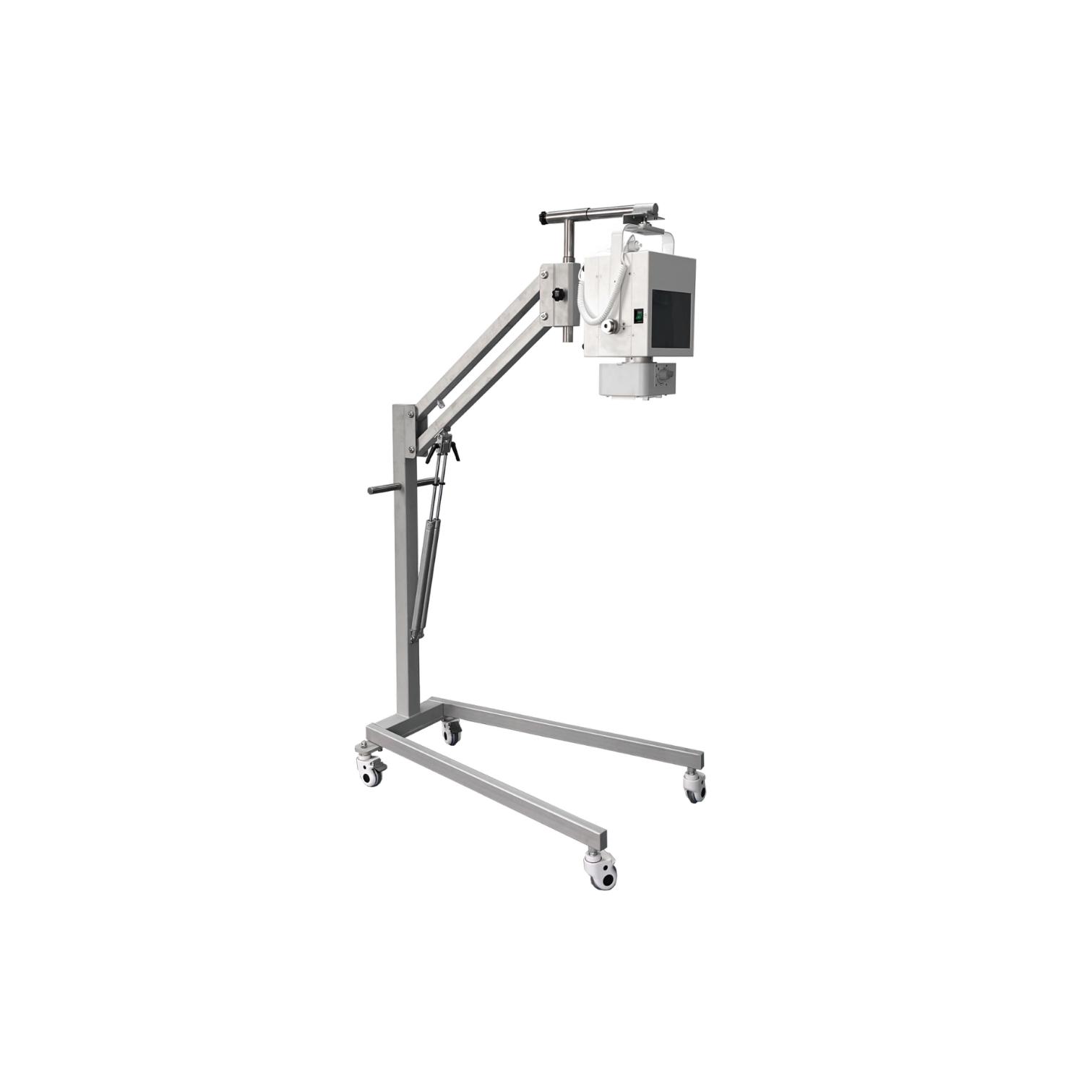
Mobile X-ray System(INP Series)
● Highly flexible: Available to put it in the flight case, easy to transfer to any place.
INP Series (Mobile Digital Radiography System)
The INP Series Mobile Digital Radiography System is designed to support fast, flexible imaging workflows in clinical environments where mobility and efficiency are critical. Its structure and operating design focus on improving positioning speed, reducing operator effort, and enabling smooth multi-area examinations.
The system allows rapid repositioning to support consecutive imaging of the chest and abdomen, helping technologists complete sequential exposures with minimal interruption and improved workflow efficiency.
Four omnidirectional swivel casters provide stable movement and precise positioning control. The unit can be guided smoothly in any direction and rotated in place, making it suitable for confined or high-density clinical spaces where maneuverability is essential.
The adjustable control panel is engineered for operational flexibility, offering both rotation and height adjustment. This allows clinicians to confirm imaging results and adjust system controls comfortably from different viewing angles during operation.
With its compact structural design and lightweight construction, the system is easy to handle during transport and positioning. The extended reach of the tube head further supports simplified one-hand positioning, allowing operators to align the system efficiently during examinations without excessive physical adjustment.
Overall, the INP Series is built to support streamlined mobile radiography workflows, combining positioning flexibility, ergonomic control design, and efficient operation in a compact mobile system suitable for a wide range of clinical environments.
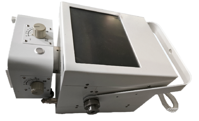
System Models Overview
| Parameter | INP-5KW-C | INP-8KW-C |
|---|---|---|
| Rated Output Power | 5 kW | 8 kW |
| Tube Current Range | 10–100 mA | 10–140 mA |
| Tube Voltage Range | 40–120 kV | 40–120 kV |
| Exposure Range (mAs) | 0.1–100 mAs | 0.2–100 mAs |
| Exposure Time | 1–2000 ms | 2–2000 ms |
| Focal Spot Size | 1.0 / 2.0 | 0.6 / 1.2 |
| Detector Type | Wireless FPD | Wireless FPD |
| Imaging Field Size | 14 × 17 inch | 14 × 17 inch |
| A/D Conversion Depth | 16-bit | 16-bit |
| Spatial Resolution | 3.4 lp/mm | 3.4 lp/mm |
| Detector Pixel Matrix | 2304 × 2800 | 2304 × 2800 |
System Functional Characteristics
-
Wireless flat panel detector (FPD) configuration supporting cable-free image acquisition and flexible positioning in clinical environments
-
Simplified exposure control interface designed for fast operation and reduced workflow steps
-
Dual control options including hand switch operation and integrated visual control console for flexible exposure triggering
-
Optimized workflow structure enabling continuous and efficient examination procedures
-
CsI direct deposition imaging technology, improving image clarity while supporting lower radiation dose requirements
-
16-bit analog-to-digital conversion system, providing enhanced grayscale depth and improved diagnostic detail
-
High-frequency inverter generator technology for stable output performance and consistent exposure quality
-
Automatic detection system for shooting distance and patient thickness, supporting optimized exposure parameter selection
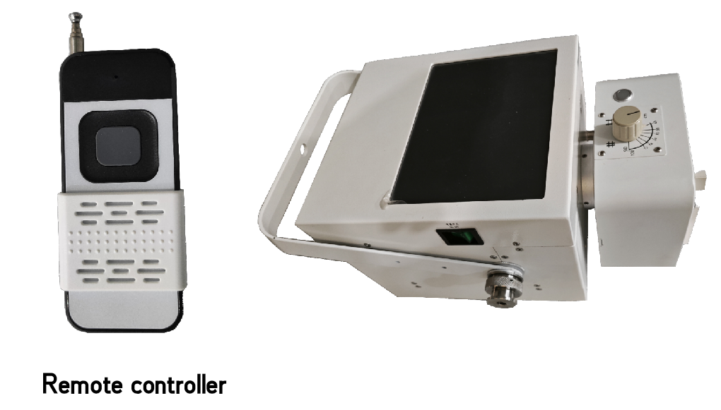
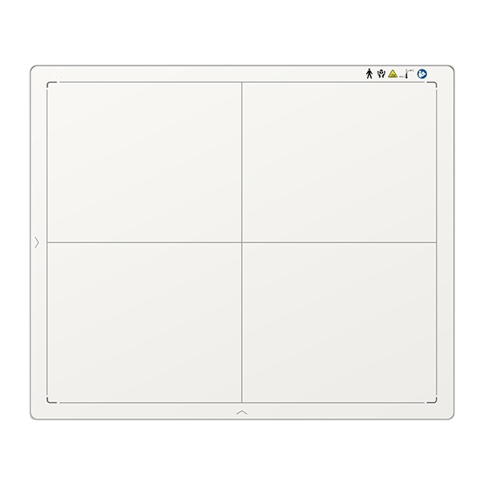
Technical Specifications
-
Detector Type: Amorphous Silicon (a-Si)
-
Scintillator Material: Cesium Iodide (CsI)
-
Active Imaging Area: 14 × 17 inch
-
Pixel Array: 2304 × 2800
-
Pixel Pitch: 150 μm
-
A/D Conversion Depth: 16-bit
-
Detection Quantum Efficiency (DQE): ≥ 70%
-
Spatial Resolution: 34 lp/cm
-
Image Acquisition Time: ≤ 2 seconds
-
Protection Rating: IPX1
Key Performance Characteristics
-
High-sensitivity amorphous silicon detector design for stable and reliable image capture performance
-
CsI scintillator layer optimized for efficient X-ray absorption and improved image sharpness with reduced radiation dose
-
Large 14 × 17 inch imaging field supporting a wide range of radiographic examinations
-
High-resolution 2304 × 2800 pixel matrix delivering detailed diagnostic image output
-
16-bit signal conversion enabling enhanced grayscale depth and improved tissue differentiation
-
High DQE (≥70%) ensuring efficient dose utilization and consistent image quality
-
Fast image acquisition cycle (≤2 seconds) supporting efficient clinical workflow
-
IPX1-rated housing structure designed for basic protection in clinical environments
Mobile Digital Radiography System — Functional Description
-
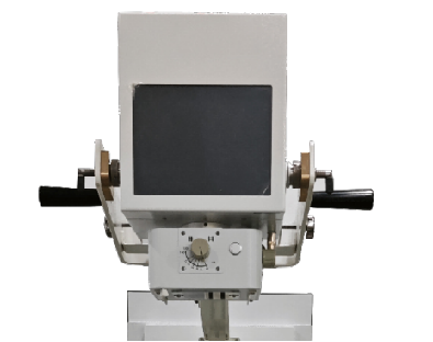
The system is designed with dual-side hand grips, allowing precise tube head positioning and improving operational efficiency during bedside examinations and polytrauma imaging scenarios, where rapid access and flexible movement are required.
-
It is equipped with a high-frequency X-ray generator available in 5 kW or 8 kW configurations, supporting up to 125 kV, 200 mA, and 400 mAs output, ensuring stable image performance and consistent diagnostic quality across different clinical conditions.
-
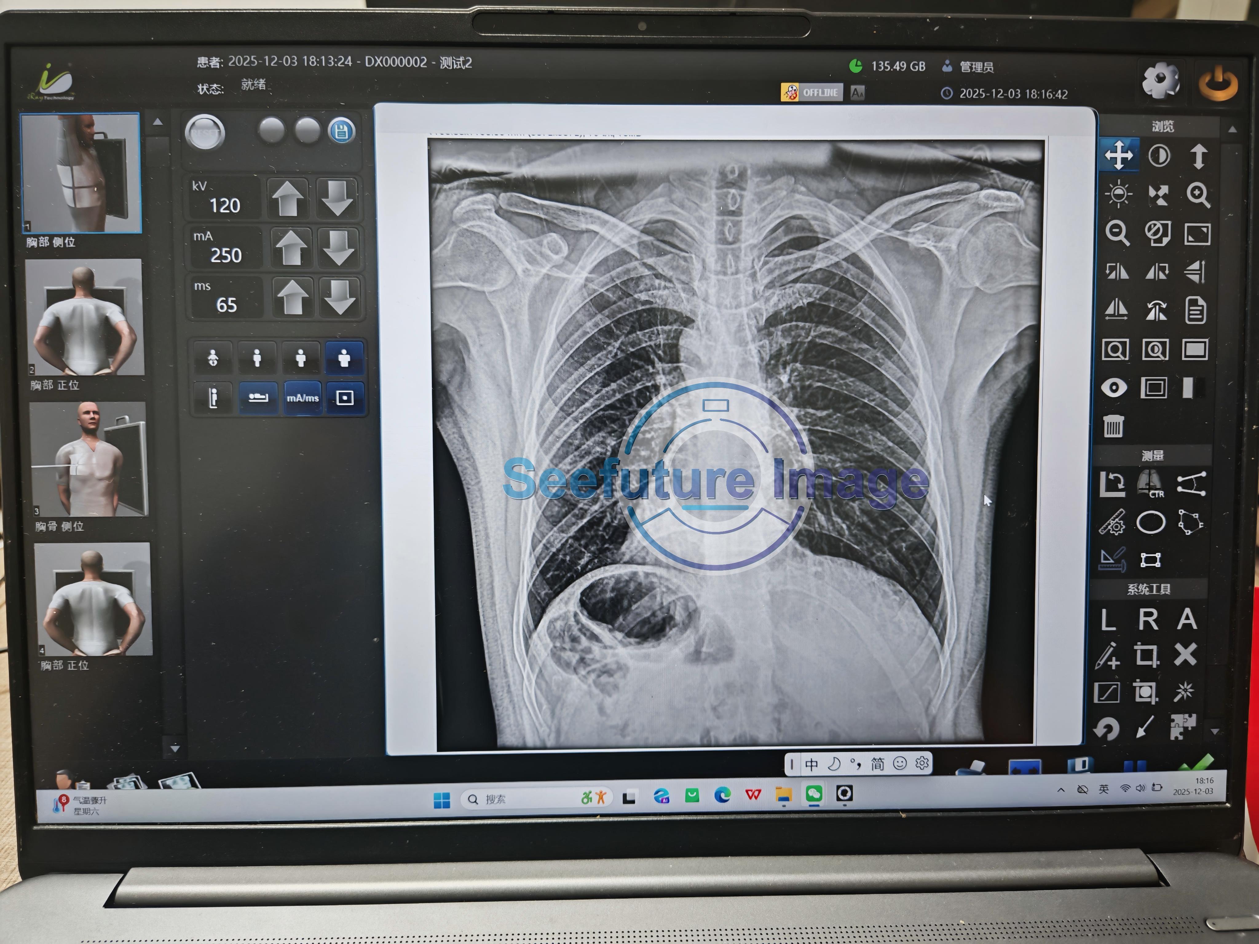
An integrated onboard computer system enables real-time image display immediately after exposure, along with fast image processing capabilities. It also supports local storage, USB data export, network-based archiving, and printing functions for streamlined workflow management.
-
The user interface is designed for simplicity and efficiency, allowing operators to configure a wide range of anatomical examination protocols. High-resolution preview images are displayed instantly prior to transmission via standard DICOM communication protocols.
-
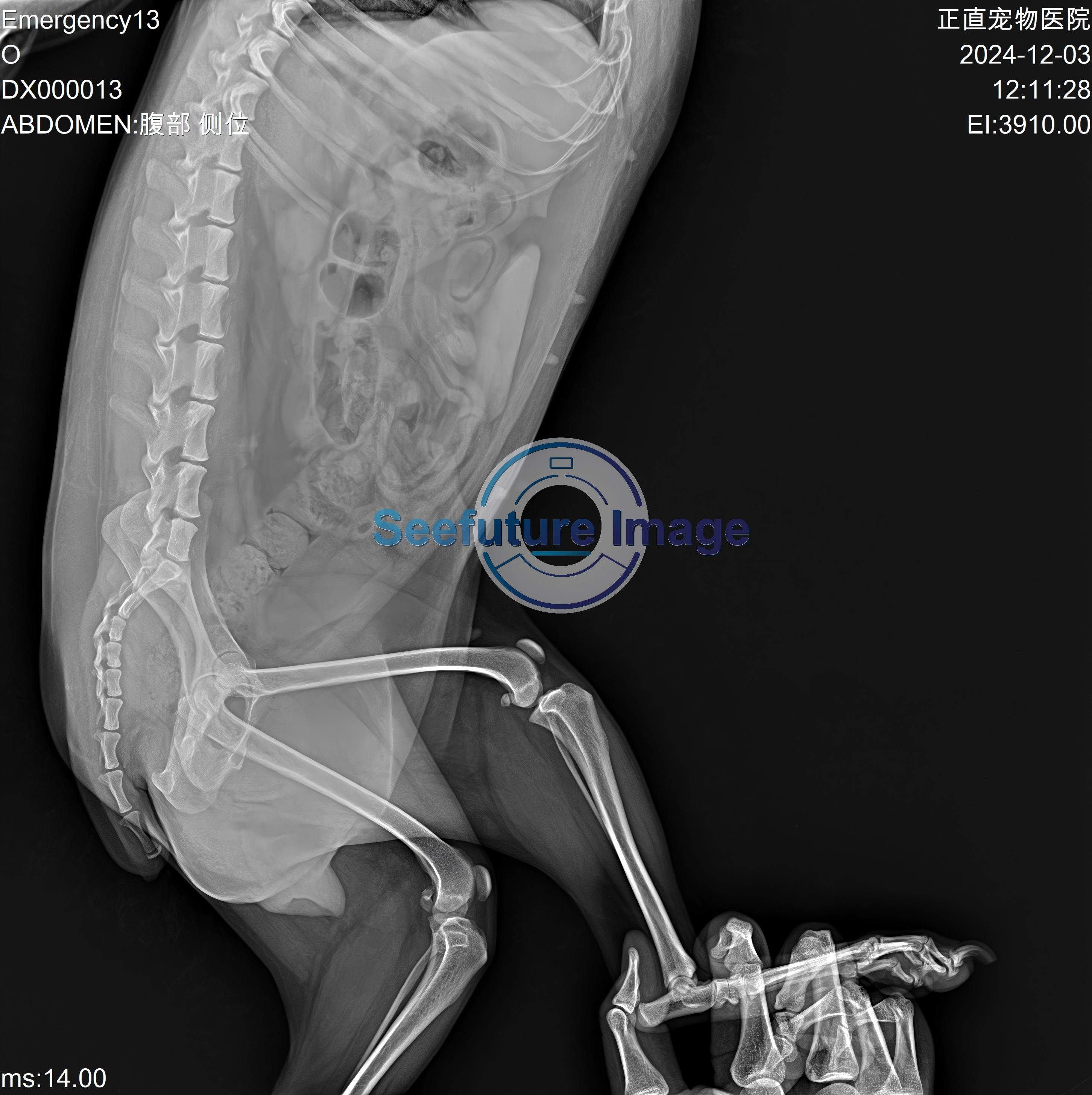
Enhanced communication and data transfer functions are integrated into the system, significantly reducing the time required for sharing imaging results within clinical workflows.
-
Multiple data connectivity options are supported, including:
-
USB external storage devices
-
CD/DVD optical storage systems
-
Ethernet wired network connection
-
Wireless (WiFi) or wired communication modes for flexible deployment in different hospital environments
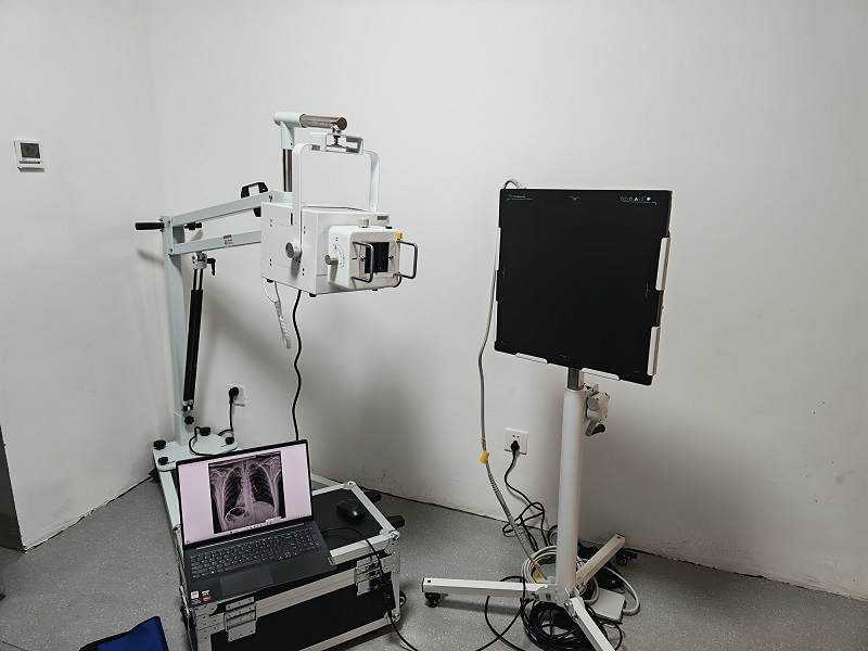
Image report sharing:




Reviews
There are no reviews yet.